What is Pollybeak Deformity?
 Deformity Turkey")
Pollybeak deformity is an aesthetic defect that typically occurs after rhinoplasty and is characterized by a noticeable fullness in the nasal profile just above the tip of the nose, i.e., in the supratip region. This condition causes the natural contour of the nasal bridge to be lost and the tip of the nose to appear lower or set back. Pollybeak deformity can develop due to factors such as insufficient cartilage and bone resection, excess soft tissue in the supratip region, thick skin structure, scar tissue developing after surgery, or prolonged edema. The deformity becomes more pronounced as the edema subsides during the healing process, and the nose may take on a profile resembling a parrot’s beak. On the other hand, while pollybeak deformity in mild cases may be confused with temporary swelling, permanent pollybeak deformity usually indicates a structural problem and often requires revision rhinoplasty to correct it.
What Does Pollybeak Deformity Look Like?
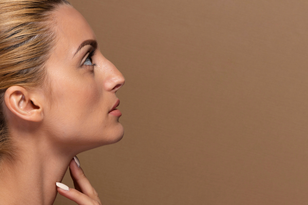
Pollybeak deformity appears as abnormal fullness in the supratip region, just above the tip of the nose when viewed in profile. This fullness disrupts the natural line of the nasal bridge, which should be straight or slightly curved, causing the tip of the nose to appear lower, set back, or drooping. As a result, the nasal profile takes on a characteristic shape resembling a parrot’s beak.
When viewed from the front, the nasal tip may not appear sufficiently defined, and the nose may give an overall heavy, coarse, or bulky impression. Supratip fullness becomes more pronounced, especially in individuals with thick skin, while the disruption of light and shadow distribution can further negatively affect the aesthetics of the nose. This appearance becomes clearer over time as the swelling subsides and is considered a significant aesthetic problem when it becomes permanent.
Who Is More Prone to Pollybeak Deformity?
Pollybeak deformity is more common in certain individuals. The main reasons for this may be related to both skin structure and post-operative care routines. In this context, pollybeak deformity is more common in the following individuals:
- Individuals with thick skin structure
- Patients with oily and dense connective tissue
- Cases where adequate support is not provided to the tip of the nose
- Individuals who develop excessive scar tissue during the postoperative healing process
- Patients who have undergone revision rhinoplasty
- Male patients
- Patients who do not adequately follow postoperative care recommendations
Is it Pollybeak Deformity or Just Swelling?
The difference between Pollybeak deformity and simple swelling is usually understood by the appearance and tissue characteristics observed after rhinoplasty. Because pollybeak deformity is a condition characterized by fullness in the supratip area, especially just above the nasal tip, or a protrusion or hump-like appearance resembling a bird’s beak, which occurs after rhinoplasty or trauma. On the other hand, simple swelling usually occurs due to temporary causes such as postoperative edema, trauma, or infection, and it diminishes and disappears over time. In this context, the swelling has a soft texture, feels sensitive or warm to the touch, and most often does not permanently alter the shape of the nose. Pollybeak deformity, however, is permanent even after the healing process is complete and usually requires surgical intervention. Consequently, the persistence of a protrusion in the nose over a long period, the hardness of the tissue, and the distinctness of the deformity may indicate pollybeak deformity rather than simple swelling.
Causes of Pollybeak Deformity
There are various causes for the development of pollybeak deformity. These causes vary from person to person and do not occur with the same severity in everyone. The causes in question are as follows:
- Excessive projection of the nasal tip cartilage: Excessive protrusion of the cartilage structure at the tip of the nose can create a bird-like appearance on the upper nasal line.
- Excess soft tissue: Excess skin and soft tissue on the tip of the nose, when combined with cartilage, can cause the deformity to become more pronounced.
- Post-rhinoplasty tissue healing irregularities: Irregular healing of tissues or scar tissue formation after surgery can trigger the Pollybeak appearance.
- Septum or upper nasal bone incompatibility: Failure to achieve adequate proportion between the upper nasal bone and the nasal tip cartilages increases the risk of deformity.
- Pre-existing anatomical abnormalities: A curved or protruding septum and cartilage structures that do not support the structure of the nasal tip pave the way for Pollybeak formation.
- Surgical technique errors: Excessive cutting of the nasal tip cartilage, incorrect shaping, or insufficient support are the main surgical causes of deformity.
Types of Pollybeak Deformity
- Soft Tissue Pollybeak Deformity
This type of Pollybeak deformity primarily occurs due to excess or thick soft tissue at the tip of the nose. Even if the nasal tip cartilages are in their normal position, excess skin and subcutaneous tissue above them creates a bird-like protrusion on the upper nasal line. It is more commonly seen in individuals with thick skin or dense subcutaneous fat tissue. Due to the soft nature of the tissue, the deformity is usually soft to the touch and can often be confused with postoperative swelling. Consequently, the treatment plan aims to reshape the soft tissue or reduce its excess.
- Cartilaginous Pollybeak Deformity
Cartilaginous pollybeak deformity occurs due to excessive projection or malformation of the cartilage structures at the tip of the nose. This condition causes a noticeable appearance resembling a bird’s beak on the upper nasal line. For this reason, cartilaginous pollybeak is usually harder and feels like cartilage rather than soft when touched. In short, the treatment performed in this procedure ensures that the cartilage is cut, shaped, or supported correctly.
- Mixed Type Pollybeak
Mixed type pollybeak is a form that combines both soft tissue excess and cartilage projection elements. Therefore, it is the most prominent and aesthetically noticeable type of deformity. This deformity causes both the shape of the tissue at the tip of the nose and the cartilage to be incompatible, making the bird beak appearance more pronounced. In this context, the treatment approach for the black type pollybeak deformity involves both thinning the soft tissue and reshaping the cartilage structure.
- Congenital Pollybeak Deformity
Congenital pollybeak deformity occurs due to congenital anatomical differences without any surgical intervention. This condition, which causes a protrusion resembling a bird’s beak at the tip of the nose, stems from genetic and structural factors. Therefore, it is usually permanent, and the individual may seek intervention for aesthetic or functional reasons. Consequently, congenital pollybeak deformity requires careful consideration of both cartilage structure and soft tissue during surgical planning to create a natural nasal contour.
How is Pollybeak Deformity Corrected?
 Deformity Turkey")
Correction of pollybeak deformity is achieved through surgical interventions planned according to the type and severity of the deformity. In cases of soft tissue-related pollybeak deformity, excess skin and subcutaneous tissue at the tip of the nose are typically thinned, and edema and scar tissue are removed, thereby reducing the prominent protrusion on the upper nasal line. In cartilaginous pollybeak deformity, the cartilage structures at the tip of the nose are reshaped, excess cartilage is removed if necessary, or supportive grafts are placed. On the other hand, in mixed types, simultaneous intervention on both soft tissue and cartilage is required; thus, both aesthetic lines are corrected and nasal functions are preserved. Finally, the same principles apply in cases of congenital Pollybeak deformity; cartilage and soft tissue are reshaped in a balanced manner, taking into account the nasal anatomy and skin thickness.
Preoperative Considerations for Pollybeak (Supratip) Deformity Surgery
There are important points to consider before pollybeak deformity surgery. Paying attention to these points helps ensure a healthier surgical process. In this context, the following are the considerations you should be aware of before pollybeak deformity surgery:
- Accurate diagnosis and deformity analysis: The type (soft tissue, cartilage, mixed) and severity of the deformity must be clearly determined before surgery.
- Assessment of patient expectations: Aesthetic goals, nose shape, and functional expectations should be discussed in detail with the patient.
- Detailed examination of nasal anatomy: Nasal tip cartilages, septal structure, upper nasal line, and skin thickness should be evaluated.
- Use of radiological and 3D imaging: When necessary, computed tomography or 3D simulations should be used to support the nasal structure and surgical plan.
- Review of previous surgery history: If rhinoplasty has been performed previously, scar tissue and existing deformities must be evaluated.
- Control of medical conditions and risk factors: Diseases and medication use that may cause bleeding, infection, or healing problems should be evaluated.
- Postoperative expectations and process information: Swelling, bruising, healing time, and the possibility of revision surgery should be clearly explained to the patient.
- Surgeon and technique selection: Choosing an experienced rhinoplasty surgeon and planning the appropriate surgical technique are critical to the success of the surgery.
Postoperative Considerations for Pollybeak (Supratip) Deformity Surgery
Just as there are points to consider before pollybeak deformity surgery, there are also points to consider after surgery. Paying attention to these points will ensure a healthier recovery process and better results. In this context, the following are the points you should pay attention to after pollybeak deformity surgery:
- Swelling and bruising control: Swelling and bruising may occur around the nose in the first few days after surgery; cold compresses and keeping the head elevated help reduce swelling.
- Nose protection and avoiding trauma: The tip of the nose and upper nasal ridge are sensitive; they should be protected from blows and hard contact.
- Nasal congestion and breathing control: If a nasal tampon or splint has been used, it should not be removed until the surgeon recommends it, and difficulty breathing is normal.
- Hygiene and risk of infection: Careful cleaning of the inside and outside of the nose is necessary, and activities that increase the risk of infection should be avoided.
- Pain and discomfort management: Painkillers and anti-inflammatory medications recommended by the surgeon should be used regularly.
- Postoperative checkups: Checkups should be attended at intervals determined by the doctor, and stitches, splints, and possible complications should be monitored.
- Restriction of physical activities: Heavy sports, exercises, and activities that put pressure on the nose should be limited for at least 4–6 weeks.
- Long-term recovery awareness: Swelling and shape changes in the nose may last for several months; the final appearance becomes clear within 6–12 months.
- Awareness of the need for revision: If the deformity does not completely resolve or if the desired shape is not achieved, a second revision surgery may be necessary; the surgeon should inform the patient about this possibility.
If you are experiencing pollybeak deformity, you can contact Dr. Hasan Duygulu, a specialist in revision rhinoplasty in Turkey, immediately.